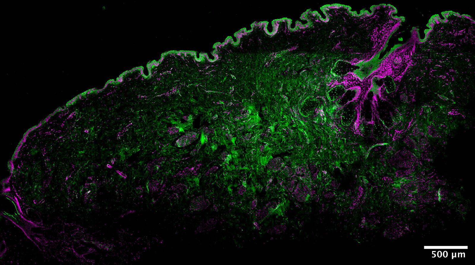
Gallery
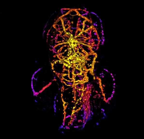
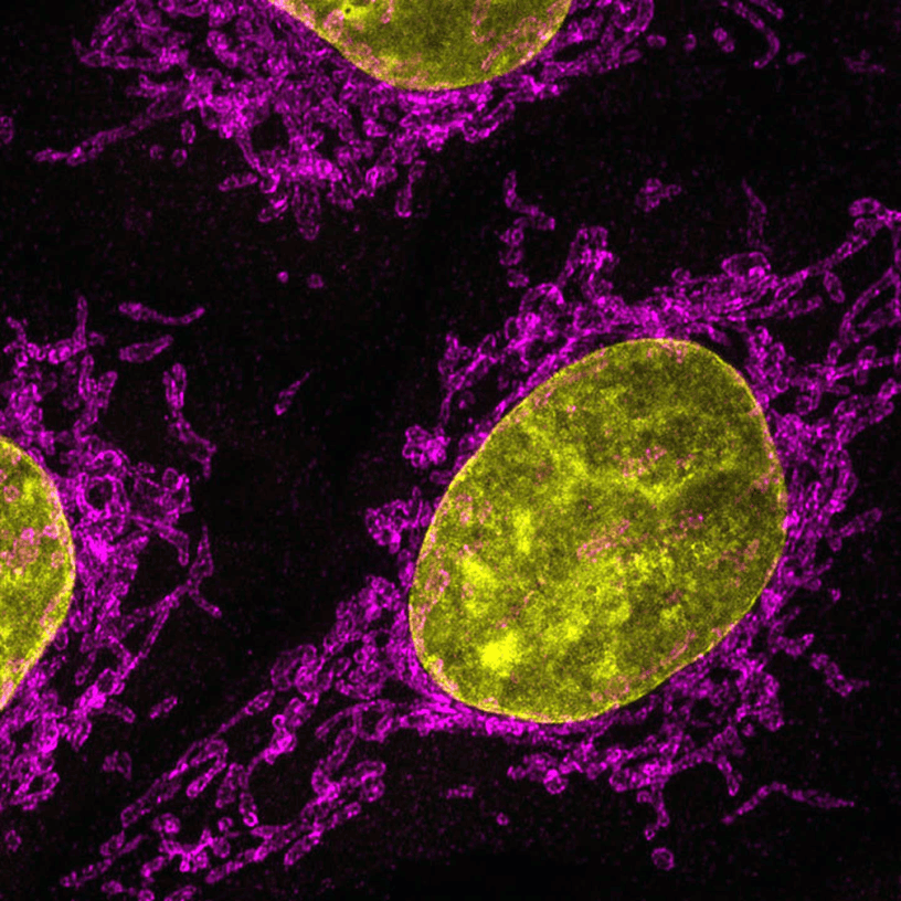
Zebrafish Embryo, Vascular System

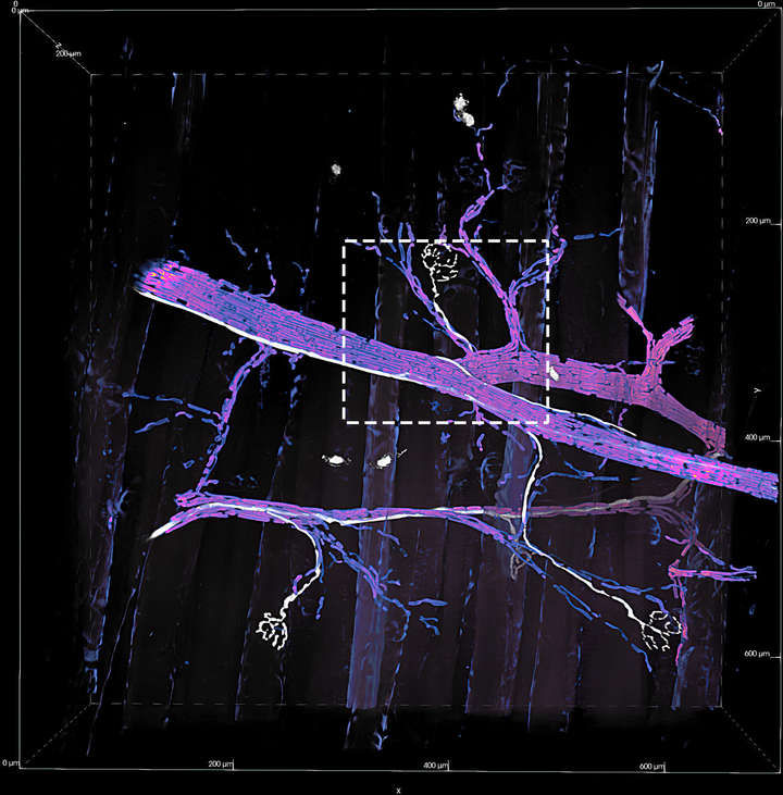
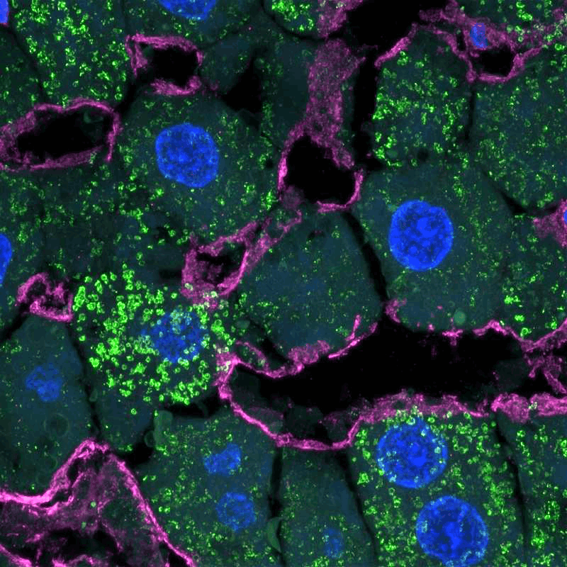
Neuromuscular Junctions

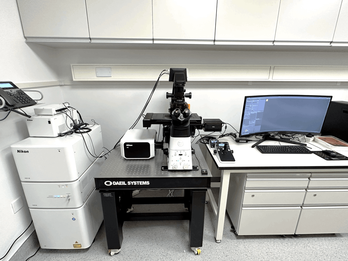
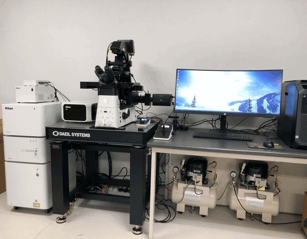
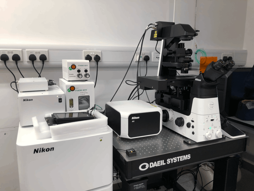
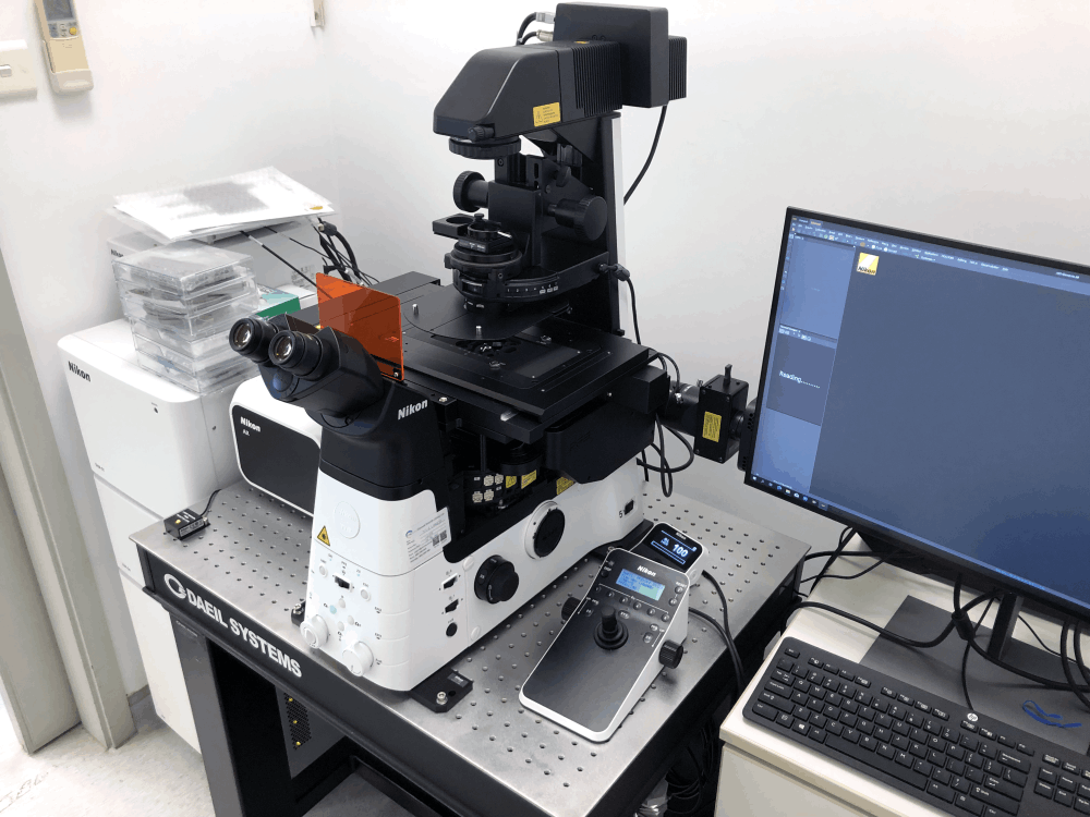
Our 10th generation point scanning confocal
- Leveraging Artificial Intelligence (AI)
- Expanding the number of colors
- Improving pixel density
- Sensitivity and speed.
Brochure >> Ask for Demo >>
With the largest field-of-view on both inverted and upright microscope stands available (25mm diagonal), more specimens fit in one FOV with more objective lens choices than ever before.
Coupled with scanning sizes up to 8192 x 8192 pixels, sampling beyond the optical diffraction limit is possible even at low magnifications with the AX/AX R.
Using lower magnifications with longer working distances and high numerical apertures enables more flexible specimen preparations to be used, while the large FOV allows simultaneous high resolution in one image. Collect more data in every image, and at faster rates.
The AX R’s high speed resonant scanning, which decreases the illumination time by more than 20x typical confocal scanning times, greatly reduces biases caused by merely acquiring images. Reducing the acquisition time also allows for extremely high-speed imaging (up to 720 fps @ 2048 x 16).
Use Mobirise website building software to create multiple sites for commercial and non-profit projects. Click on the video preview in this block to add the link to your video. Make sure that your video is not private. You can add a description below the video. If you want to hide some of the text fields, open the Block parameters, and uncheck relevant options.
With a full 25mm FOV, up to 8192 x 8192 pixels, and the capability for supravideo frame rates, the AX/AX R allows for spectacular imaging with high resolution, at both low and high magnifications.
The entire range of a whole organism or system biology down to intracellular imaging is achievable on one instrument.
Autosignal.ai
adjust to the best illumination and detection settings automatically
Denoise.ai
Remove the shot noise component, improve the image quality, and assist in downstream segmentation
Segment.ai
By tracing features of interest and training these compared to the underlying image, the neural network can learn and apply segmentation to similar images, recognizing features previously only identifiable by painstaking manual tracing approaches.
*Photos and Videos courtesy of Nikon official website
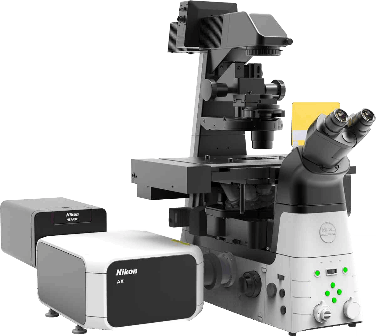
AX Confocal with NSPARC
Low noise Nikon Spatial Array Confocal (NSPARC) detector, resulting in high sensitivity, high signal-to-noise, and possibility of live-cell assay.
Learn more>>
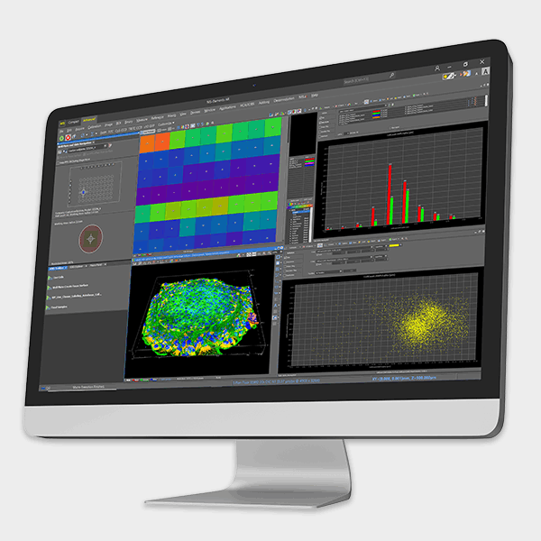
With Nikon microscope and software NIS-Elements, our high content imaging streamlines high-speed imaging and simple operations. Nikon software provides a dedicated interface for high content acquisition and analysis routines.
Without the need to adjust the focusing method, wavelengths, filters, and camera settings, it is easier to process alongside your research quickly and effortlessly.

A study of living cells using microscopy to obtain a better understanding of biological function through the study of cellular dynamics.
Fluorescence itself is a form of luminescence that results from matter emitting light of a certain wavelength after absorbing electromagnetic radiation. Fluorescence Imaging help visualize biological processes taking place in a living organism.
Is the study of the nervous system. The scope of neuroscience has broadened over time to include different approaches, such as optogenetics, a biological technique to control the activity of neurons or other cell types with light. This is achieved by the expression of light-sensitive ion channels, pumps, or enzymes specifically in the target cells.
Phone & WhatsApp:(+852) 2784 0630
Email: sales@ctschina.com.hk
Working Hour: 0930-1700
© Copyright 2025 Chinetek Scientific (China) Limited - All Rights Reserved

Drag & Drop Website Builder